Source: Doctors for Covid Ethics
Alternate mechanisms of mRNA vaccine toxicity: which one is the main culprit?
by Michael Palmer, M.D.
May 3, 2023
This paper summarizes the mode of action of mRNA vaccines, as well as three potential pathogenetic mechanisms that may account for the toxicity observed with the mRNA vaccines against COVID-19, namely: chemical toxicity of lipid nanoparticles, direct toxicity of the spike protein, whose expression is induced by the vaccines, and the destructive effects of the immune response to the spike protein. The case is made that of these mechanism the third is likely the most important one. If this conclusion is correct, then essentially the same level of toxicity must be expected with future mRNA vaccines against any other pathogenic microbes.
1. How mRNA vaccines provoke immune attack on our own cells and tissues
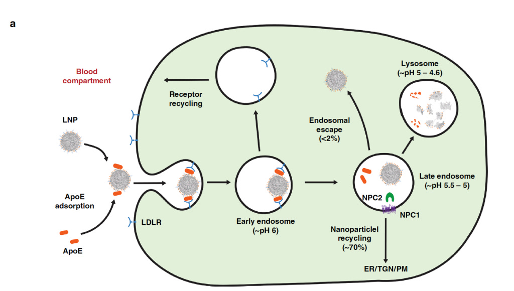
mRNA vaccine particles have two components—the mRNA itself, and the lipid mixture which encloses it. The lipids protect the mRNA while in transit, and they facilitate its cellular uptake.
Once within the cell, the mRNA binds to ribosomes—the little grey blobs in the cartoon—which read the sequence of the mRNA and assemble the protein accordingly. In case of the COVID-19 vaccines, the product is the spike protein of the SARS-CoV-2 virus.
The spike protein will be taken to the cell surface, where it may be bound by antibodies. Those bound antibodies will activate the complement system, a cascade of serum proteins which culminates in the formation of a membrane attack complex. Such complexes create large holes in the cell membrane, ultimately killing the cell.
Another pathway to immune attack begins with the fragmentation of some spike protein molecules within the cell. The fragments are again taken to the cell surface, where they are recognized by cytotoxic T-lymphocytes (T-killer cells). Recognition causes them to attack and kill the infected cell.
The above assumes that we already have antibodies which recognize the spike protein or its fragments. How do these arise in the first place? Without going into details, we note that their induction requires some measure of cell damage and cell death, brought on for example by T-killer cells. The antibody response is then induced by the debris of those dead cells.
1.1. Sure, that looks pretty bad, but aren’t mRNA vaccines just like live virus vaccines?
It is true that the same two pathways also operate in the immune defence against natural viruses and live virus vaccines. However, the devil is in the details.
1.2. Three key differences between live virus vaccines and mRNA vaccines
Live virus vaccines mRNA vaccines Replication inside the host cell yes no Vaccine particles contain protein antigens yes no Vaccine particles infect blood vessel walls no yes
We will see presently why these three differences are important.
1.3. The time course of viral load and immune response in a virus infection or live virus vaccination
If we are infected with a natural virus or inoculated with a live virus vaccine, the initial viral load is small. In the absence of prior immunity, the virus will replicate for a good while, and the viral load will increase until it is checked by the onset of the immune response. By the time the immune response reaches its peak, the viral load will typically already have declined, and the fight against the infection will be at the “mop-up” stage.
A secondary infection will trigger a memory response, which curbs the multiplication of the virus early on. Therefore, the peak of the viral load will remain much lower, and the infection will often not even be clinically apparent—we are “immune” to it. Again, any small-scale infection that does occur will be largely gone by the time the immune response attains its peak. Thus, neither with the primary infection nor with a secondary one will peak viral load and peak immune response clash head-on. This limits the intensity of inflammation.
1.4. Repeat injections of mRNA vaccines will clash with existing immunity
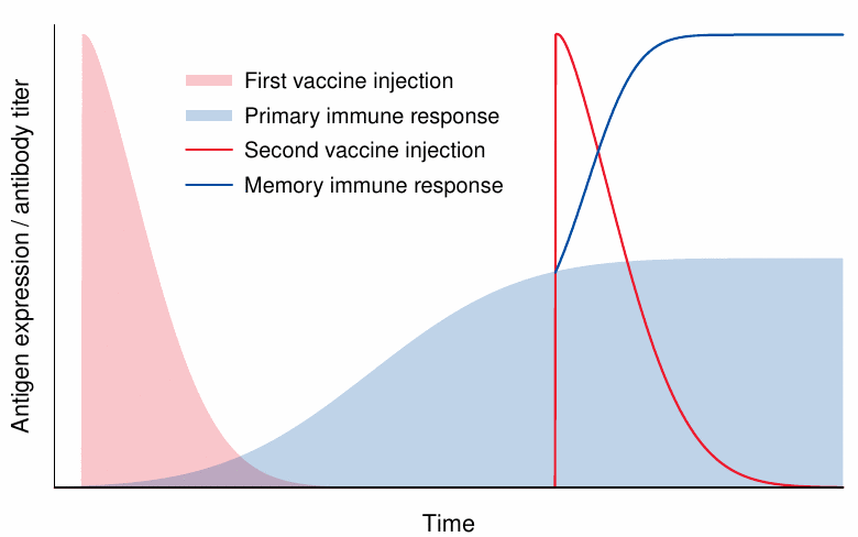
We now come to the first disparity between viruses and mRNA vaccines—namely, that unlike viruses mRNA vaccines do not replicate. This makes it necessary to inject the full amount of vaccine particles all at once and every time.
The expression of the antigen is supposed to decline on a time scale of days. If we assume this to be the case, and also that no immunity yet exists, we may again avoid a clash between peak antigen expression and peak immune response. However, with a repeat injection, and also in case of natural immunity due to a previous infection with the virus, we must expect antigen expression to clash head-on with an intense immune response, resulting in accordingly intense inflammation. Thus, both acute side effects and long-term ones such as autoimmune disorders will become more likely after the second shot.
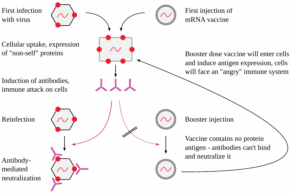
1.5. Unlike viruses, mRNA vaccine particles aren’t recognized by antibodies
The concern about the head-on clash between vaccine antigen expression and immune response is compounded by the second disparity between viruses and mRNA vaccines—namely, that the mRNA vaccine particles do not contain any copies of the encoded protein antigen on their surfaces.
The presence of protein antigens on virus particles means that these can be bound by antibodies that are already present, which will prevent those virus particles from infecting our body cells. Even though some particles may still manage to get through, the antibodies will at least mitigate the infection.
In contrast, mRNA vaccine particles cannot be stopped by antibodies at all, for the simple reason that they contain only the nucleic acid blueprint for the protein but not the actual protein itself. Therefore, these particles will be taken up by our body cells regardless of immunity. Any immunity already present will then be directed against those unlucky cells.
Thus, in a nutshell, with real viruses existing immunity will inhibit cell damage and inflammation, while with mRNA vaccines existing immunity will make things worse.
1.6. Summary: the inherent flaws of mRNA vaccines
High particle load clashes with intense immune response
Particles “fly under the radar” of antibody surveillance before entering cells, directing an “angry” immune system against those cells
Antigen expression in cells of blood vessel walls causes destruction of vessels, with activation of blood clotting
The first two points promote intense inflammation, with severe tissue destruction and the risk of triggering autoimmunity. Damage to blood vessels poses grave risks as well; this is illustrated by the severe disease caused by those natural viruses which do infect blood vessel walls. Examples are the Ebola, Marburg, and Dengue viruses.
2. Adverse event severity after repeated vaccine injection
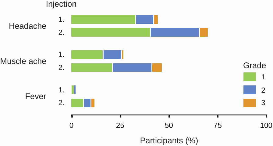
2.1. Increased severity of adverse events in teens after the second shot of Moderna vaccine
This figure is adapted from Ali et al. [1], the “clinical trial” on the Moderna vaccine in teenagers. We see a general increase in severity after the second injection, particularly with grade 2 and 3 side effects. Grade 3 fever means between 39 and 40 degrees centigrade. Such high fever signals a potential for severe inflammation and is normally considered unacceptable in a vaccine candidate.
2.2. Cardiac symptoms in teens after first and second injections with the Pfizer vaccine
The data in this figure are from Chiu et al. [2], who followed approximately 5000 injected teenagers. We see a clearly increased incidence of symptoms after the second injection.
The cardiac symptoms monitored in this study are comparatively “mild”, but they are very frequent, leading us to expect that more severe cardiac consequences will occur as well. Many of these symptomatic vaccinees likely incurred some degree of inflammation in the heart.
2.3. VAERS reports of myocarditis after the first and the second dose
Of course, we already know that vaccine-induced myocarditis is not rare. This figure (adapted from Oster et al. [3]) shows that with myocarditis, too, the incidence is significantly higher after the second injection than the first. A study from Basel in Switzerland suggests an even higher incidence after the third injection [4].
2.4. Days to death by age and dose (VAERS, up to 2021-11-23)
This analysis, carried out by Craig Pardekooper of “How bad is my batch” fame [5], suggests that the story is a little more complicated. Apparently, there are more delayed deaths, but fewer early ones after the second dose.
The total numbers of deaths in both panels are quite similar. However, it stands to reason that the likelihood of the connection being made between an injection and a death or other severe adverse event decreases with time; or in other words, that the under-reporting bias will increase with time. Therefore, delayed deaths are likely under-represented relative to the early ones. This suggests that overall mortality will be significantly greater after the second dose.
3. Alternate mechanisms of vaccine injury
Chemical toxicity of lipid nanoparticles
Spike protein toxicity
Immune response to spike protein as a foreign antigen
Induction of genetic mutations by the mRNA and by contaminating DNA
In the following, we will look briefly at the first three mechanisms, and then consider which one is most likely predominant. The fourth mechanism was discussed in a previous article [6].
3.1. Cationic lipids are highly inflammatory
This figure is adapted from a paper by Ndeupen et al. [7], who injected mice with lipid mixtures similar to those used in the COVID-19 mRNA vaccines. The left panel shows excised skin patches from mice that had been injected with buffer (PBS) only, with an inert lipid mixture (nNLP), or with a lipid mixture containing cationic lipids (iLNP). In the latter case, the skin patches are bloodshot, indicating inflammation. The plots to the right show that inflammatory cells, in particular neutrophil granulocytes, are increased in this case as well.
3.2. Cationic lipids induce reactive oxygen species, which can induce apoptosis (programmed cell death)
Cationic lipids can also induce programmed cell death (apoptosis), which can be detected by DNA fragmentation, as shown in this electrophoresis experiment. DNA fragments are here separated according to size—the further from the origin on the left, the smaller they are. In cell cultures, this effect can be suppressed by the addition of a reducing agent (N-acetylcysteine), which indicates that it is mediated by reactive oxygen species (ROS).
The induction of ROS by cationic lipids has been confirmed in multiple studies, and it poses a risk of DNA damage even if the threshold of outright apoptosis is not reached. This risk is further substantiated by some very fragmentary data on genetic damage in red blood cell precursors, which were submitted by Moderna to the European Medicines Agency [8].
3.3. Toxic activities of spike protein
Spike protein fragment S1 can be cleaved off cell surfaces and enter the bloodstream
S1 fragment binds and inhibits angiotensin converting enzyme (ACE2)
ACE2’s substrate, angiotensin II, accumulates and causes
elevated blood pressure
activation of blood clotting
increased inflammation
Intracellular spike protein inhibits DNA repair
The spike protein itself also has toxic activity, which may be mediated by inhibition of ACE2 [9,10] and potentially also by the inhibition of DNA repair [11], which may compound the mutagenic risks posed by the RNA and contaminating DNA [6] as well as the cationic lipids.
3.4. Immune pathology after vaccination (“lymphocyte amok”)
This slide shows normal heart muscle tissue for comparison, and heart muscle tissue affected by vaccine-induced myocarditis, as evident from the cluster of lymphocytes in the middle. This image is from an autopsy case reported by pathologist Prof. Arne Burkhardt, who has used the term “lymphocyte amok” to sum up his findings in many organs and in many patients who had died after receiving gene-based COVID-19 vaccines.
3.5. Healthy lung tissue, and clusters of lymphocytes in the lung of a vaccine victim
This picture, likewise from Prof. Burkhardt, shows lymphocytic inflammation of the lung. The normally delicate alveolar septa of the lung tissue are thickened by scar tissue, and the air-filled spaces (white) are compressed.
The dark blue dots in the left panel are the cell nuclei of the regular cells within the tissue. Some of the blue dots in the right panel are of the same nature. However, the large cluster within the top left quadrant is an accumulation of invading lymphocytes.
3.6. Vasculitis of small blood vessels in the brain
Blood vessels of all sizes are prominently affected by lymphocytic inflammation. This slide (again by Prof. Burkhardt) shows two inflamed small vessels within brain tissue. On the right, the inflammatory lesions have caused the blood within the vessel to clot.
Lymphocytes are also seen in the surrounding brain tissue itself, indicating that inflammation is not limited to the blood vessels.
3.7. Spike protein expression in a case of vaccine-induced encephalitis
The causal connection between vaccine injection and inflammation can be demonstrated by immunohistochemistry. In this method, the specificity of antibodies for a given protein of interest—such as in our case the spike protein—is used for its sensitive and selective detection in tissue sections. Several steps downstream of antibody binding, a brown pigment is generated that shows the presence and distribution of the target protein.
This slide shows brain sections from a patient who died several weeks after receiving his most recent COVID-19 vaccine injection [12]. The conventional (HE) stain on the right shows a dead nerve cell (1) and inflammatory cells including lymphocytes (2). The diagnosis made was necrotizing encephalitis, i.e. brain inflammation with cell/tissue death.
In the left panel, we see that spike protein is expressed by several cells in a sample of brain tissue, as evidenced by the deposition of brown pigment atop those cells. The middle panel shows that the nucleocapsid—another protein of the SARS-CoV-2 virus—is not expressed. The vaccine encodes only the spike, whereas virus-infected cells should express all viral proteins, also including the nucleocapsid. Therefore, the spike protein which is detected must have been induced by the vaccine. This fits with the clinical history in this case, which included three vaccinations but no diagnosis of the virus infection.
3.8. Myocarditis with complement deposits and primarily granulocytic infiltrates
This case of myocarditis with sudden death was reported by Choi et al. [13]. Five days after the first dose of Pfizer vaccine, the patient developed chest pain, and he died seven hours later. Complement factor C4 deposits are shown by immunohistochemistry in panel C. Cellular infiltrates predominantly with neutrophil granulocytes are seen in panels A and B. The horizontal red stripes in panel B are contraction band necroses, which are known to occur after intracellular calcium excess.
Complement activation will result in membrane permeabilization (see Section 1), which accounts for the calcium excess and the contraction band necroses. Complement activation will also release chemotactic factors that will attract granulocytes to the scene. This case therefore exemplifies the antibody/complement-mediated pathway of immune attack on cells which express the mRNA vaccine-encoded antigen. Most likely, the unfortunate patient already had strong natural immunity to the SARS-CoV-2 virus.
4. Which of the three mechanisms is the dominant one?
Mechanism Expected effect of 2nd dose Comment Lipid toxicity no change toxicity of adenovirus-based vaccines? Direct spike protein toxicity reduced responsible for rapid effects of first dose? Immune response to spike protein increased completely general—must be expected with all future mRNA vaccines
One key consideration for identifying the dominant mechanism of mRNA vaccine toxicity is how each of them should change in intensity with the second injection.
With the cationic lipids, one might expect similar levels of acute toxicity after each application, although some degree of cumulative acute toxicity is conceivable. We note, however, that the spectrum of adverse events observed with adenovirus-based COVID-19 vaccines is very similar to that of the mRNA vaccines, even though the virus-based vaccines don’t contain any cationic lipids. This finding suggests that the common toxic principle of both vaccine types is the spike protein—either its direct toxicity, or the immune response to it.
Direct spike protein toxicity should be mitigated by specific antibodies to it, just as diphtheria and tetanus are mitigated by antibodies to their respective protein toxins. Thus, we should expect that the antibodies induced by the first vaccine injection should reduce the severity of adverse events that follow the second injection. However, as noted in Section 2, the severity of side effects tends to increase with repeated application of the vaccine. The one exception were early deaths, which were indeed more frequent after the first injection than the second one (see Section 2.4). The same trend was also found by Craig Pardekooper with non-deadly adverse events. It seems possible, therefore, that many adverse events which occur early on after the first injection are indeed related to the direct toxicity of spike protein, which manifests itself in the time window between the injection and the induction of antibodies.
A key role of the immune response to spike protein is supported by the following arguments:
it follows from the theoretical considerations spelled out in Section 1;
it agrees with the predominant trend of increased adverse event incidence and severity after repeated vaccine injection;
it accounts for the histopathological findings of intense inflammation and infiltration with lymphocytes as well as other immune cells, which are observed near foci of spike protein expression.
Why does it matter which of the pathogenetic mechanisms is predominant? There are plans to convert existing vaccines, including childhood vaccines, to mRNA technology. If direct toxicity of the SARS-CoV-2 spike protein were mainly responsible for the adverse events caused by the COVID-19 mRNA vaccines, then future mRNA vaccines might be more benign, as long as the antigenic proteins which they encode are less toxic than the SARS-CoV-2 spike protein.
On the other hand, every mRNA vaccine will induce an immune response in the same manner as the COVID-19 mRNA vaccines. Therefore, if that immune response were mainly responsible for toxicity, then we must expect similarly catastrophic outcomes with all future mRNA vaccines. The arguments presented in this study indicate that this is the most likely case.
The contributions of the other two mechanisms cannot be ignored. Direct toxicity appears to significantly contribute to early toxicity after the first injection. Cationic lipid toxicity cannot be dismissed either, for the following reasons:
almost no safety studies were conducted on these substances during the dysfunctional approval processes of the COVID-19 vaccines, but the rudimentary ones which were performed gave clear indications of toxicity;
the induction of reactive oxygen species (ROS) by cationic lipids will cause DNA damage. This damage will stay behind even after the lipids themselves have been eliminated, which means that toxicity will be cumulative;
since cationic lipids are a necessary ingredient of all mRNA vaccines, their toxicity will accumulate across all doses of all mRNA vaccines, rather than just across all doses of a single one.
In conclusion, the whole of the evidence indicates that all three mechanisms of toxicity must be taken seriously. At the same time, it points to a key role of immune-mediated toxicity, which results from the fundamental flaws of the mRNA vaccine technology and must be expected to occur with future mRNA vaccines directed against any and all viruses or other pathogenic microbes.
References
Ali, K. et al. (2021) Evaluation of mRNA-1273 SARS-CoV-2 Vaccine in Adolescents. N. Engl. J. Med. DOI:10.1056/NEJMoa2109522
Chiu, S. et al. (2023) Changes of ECG parameters after BNT162b2 vaccine in the senior high school students. Eur. J. Pediatr. 182:1155-1162
Oster, M.E. et al. (2022) Myocarditis Cases Reported After mRNA-Based COVID-19 Vaccination in the US From December 2020 to August 2021. JAMA 327:331-340
Le Pessec, G. et al. (2022) Significant incidence of myocarditis after 3rd dose of RNA vaccine anti-COVID messenger 19.
Pardekooper, C. (2023) How Bad is My Batch? Batch codes and associated deaths, disabilities and illnesses for Covid 19 Vaccines.
Palmer, M. and Gilthorpe, J. (2023) COVID-19 mRNA vaccines contain excessive quantities of bacterial DNA: evidence and implications.
Ndeupen, S. et al. (2021) The mRNA-LNP platform’s lipid nanoparticle component used in preclinical vaccine studies is highly inflammatory. iScience 24:103479
Anonymous, (2021) EMA Assessment report: COVID-19 Vaccine Moderna.
Marik, P. (2021) An overview of the MATH+, I-MASK+ and I-RECOVER Protocols (A Guide to the Management of COVID-19).
Angeli, F. et al. (2022) COVID-19, vaccines and deficiency of ACE2 and other angiotensinases. Closing the loop on the “Spike effect”. Eur. J. Intern. Med. 103:23-28
Jiang, H. and Mei, Y. (2021) SARS-CoV-2 Spike Impairs DNA Damage Repair and Inhibits V(D)J Recombination In Vitro. Viruses 13:2056
Mörz, M. (2022) A Case Report: Multifocal Necrotizing Encephalitis and Myocarditis after BNT162b2 mRNA Vaccination against Covid-19. Vaccines 10:2022060308
Choi, S. et al. (2021) Myocarditis-induced Sudden Death after BNT162b2 mRNA COVID-19 Vaccination in Korea: Case Report Focusing on Histopathological Findings. J. Korean Med. Sci. 36:e286
The Shannon Joy Show
The Vaccines Are BioWeapons & If We Want To WIN We Must Acknowledge This w/ Sasha Latypova
Sasha identifies the nature of this 21st century war, the players good and bad AND the ways we will WIN it through widespread, PEACEFUL non-compliance. But the FIRST step is acknowledging the fact itself.
Source: The Daily Clout
Report 74: Lipid Nanoparticles Corrupt Nature
by A War Room/DailyClout Pfizer Documents Analysis Project Volunteer report, edited by Chris Flowers, M.D.
June 5, 2023
INTRODUCTION
This report delves into the history of lipid nanoparticles, as well as their use by pharmaceutical companies as a casing for mRNA in the COVID vaccines. It concludes that the use of the lipid nanoparticles to deliver mRNA causes widespread and significant harms to the human body.
Without the lipid nanoparticles, mRNA cannot enter human cells, and the body would recognize the mRNA as a foreign and destroy it. In order to allow the mRNA to travel throughout the body, the mRNA is encased in lipid nanoparticles.
The lipid nanoparticles are synthetic fat molecules that mimic natural fat molecules. This means that they are not recognized by the body as a threat and are not destroyed by immune response as a foreign invader, which allows them to enter and release their mRNA cargo inside of cells. The mRNA then takes over and tricks the cells into synthesizing its foreign protein.
LIPID NANOPARTICLES CORRUPT NATURE
Lipid nanoparticles (LNPs) are synthetic lipids developed in the laboratory, and the technology has many unknowns. The technology is so new that there are no long-term studies on their health impacts. However, studies done before Pfizer was granted Emergency Use Authorization (EUA) revealed LNPs travel everywhere in the body and collect in most, if not all, organs. Pfizer told the world its mRNA gene therapy drug stays in the arm into which it is injected, even though the Pharma giant knew from its own biodistribution study that that was a lie. [“A Tissue Distribution Study of a [3H]-Labelled Lipid Nanoparticle-mRNA Formulation Containing ALC-0315 and ALC-0159 Following Intramuscular Administration in Wistar Han Rats,” https://www.phmpt.org/wp-content/uploads/2022/03/125742_S1_M4_4223_185350.pdf, pp. 23-27.]
This report will bring facts and give clarity and motivation for readers to research for themselves, decide if they are being told the truth, and then determine if they really want to take an experimental gene therapy drug.
NANOPARTICLES OF THE PAST
In nature, nanoparticles have always existed. Carbon nanotubes, discovered in the coating of pottery from India dating 600-300 BC, enabled it to last for centuries. However, there is no way to know if the nanotubes were there by accident or not. Also found circa 900 AD, Damascus steel contained cementite nanowires, and the origins and how they were made are unknown. (https://www.indiatvnews.com/news/india/oldest-known-human-made-nanostructures-dated-to-600-bc-discovered-in-tamil-nadu-ancient-artifacts-666406) (TEM image of cementite nanowires in Damascus steel: https://www.researchgate.net/figure/TEM-image-of-cementite-nanowires-in-Damascus-Steel-a-and-b-Carbon-nanotubes-in_fig2_258434890)
In the eighth-century Mesopotamia, which is modern-day Iraq, artisans were known for creating a glittering effect on a pot’s surface known as Lusterware. The process involved applying a mixture that included metal nanoparticles applied on previously glazed items, giving it an iridescent, metallic luster. (https://www.thoughtco.com/what-is-lustreware-171559)
Dawn of Synthetics
Modern-day nanotechnology first began in 1833 when the word “polymer” was introduced by a world-renowned Swedish chemist, Jöns Jakob Berzelius.
A year later, he introduced the term “isomer,” meaning substances with identical but differing properties. In other words, the substances have the same number and types of atoms but differ because the arrangement of the atoms makes them have different chemical structures. (https://www.britannica.com/biography/Jons-Jacob-Berzelius/Organic-chemistry)
This later helped launch an explosion of new synthetic polymers in the 1930s and 1940s, especially after World War II, including polyvinyl chloride (PVC), polyurethane (PU), nylon fibers, neoprene (synthetic rubber), polytetrafluoroethylene (PTFE or Teflon), and polystyrene (PS). Suddenly, synthetic fibers, films, plastics, rubbers, coatings, and adhesives were everywhere. (https://www.azom.com/article.aspx?ArticleID=19088, https://www.polyplastics.com/en/pavilion/beginners/01-05.html, https://www.globalspec.com/reference/32095/203279/chapter-twelve-synthetic-petroleum-based-polymers, https://theconversation.com/if-plastic-comes-from-oil-and-gas-which-come-originally-from-plants-why-isnt-it-biodegradable-179634, and https://www.ifc.org/wps/wcm/connect/f0879328-5764-4004-a5c4-1ce906701170/Final%2B-%2BPetroleum-based%2BPolymers%2BMnfg.pdf?MOD=AJPERES&CVID=jkD2Egn&id=13231530.)
But these petroleum-sourced polymers contrast with biopolymers. Biopolymers are polymers from sources in nature and can be produced chemically from biological materials or are made by living organisms.
THE DISCOVERY OF LIPOSOMES
In 1965, Alec D. Bangham, a British biophysicist discovered liposomes (derived from two Greek words: lipo (“fat”) and soma (“body”)),
and composed primarily of phospholipids, which are phosphorus-containing complex lipids and closed lipid bi-layer vesicles.
Bangham found that liposomes could be made spontaneously in water. These were the earliest version of lipid nanoparticles. Liposomes became the most widely studied and recognized drug delivery platform due to their biocompatibility and biodegradability, which resulted in minimal adverse reactions. They are currently used in skin care products and in nutritional supplements as well. (https://www.thelancet.com/pdfs/journals/lancet/PIIS0140-6736(10)60950-6.pdf and https://www.sciencedirect.com/science/article/abs/pii/S0924224401000449)
Liposomes used for drug treatments have had challenges, including poor stability and storage, and they are affected by pH and temperature. They have a low encapsulation rate due to a leakage problem with water-soluble drugs. Now, the use of PEGylated liposomes helps to improve encapsulation efficiencies and the size of particles, giving them a “stealth” effect, which increases their circulation time in the body. Research is ongoing. (https://pubmed.ncbi.nlm.nih.gov/32650041/ and https://pubmed.ncbi.nlm.nih.gov/32431154/)
LIPOSOMES AND LIPID NANOPARTICLES
Liposomes are hollow with one or more rings of lipid bilayers and are spherical vesicles surrounding a watery core, used as a drug carrier, made mainly from phospholipids and other physiologic lipids.
LNPs are made with lipid, or fat, components other than phospholipids. They are novel synthetic and semi-synthetic lipids that have greater stability because reactants act very slowly. LNPs have a more rigid form and structure, and this gives them significant advantages over liposomes.
The term ‘lipid nanoparticle’ originated in the 1990s. Big Pharma’s seems to have its sights set on revolutionizing healthcare using LNPs. LNPs are tiny fat balls, measured in nanometers, at a size of less than 200nm. A nanometer is one billionth of a meter. In comparison, the width of a human hair is 80,000nm to 100,000nm.
The science behind LNPs is an idea taken from nature. They are created using a micelle-like structure that holds drug molecules in a non-watery core surrounded and protected by a double-layer membrane of different lipids. With gene therapy, it is the encapsulated artificial pseudouridine (Pfizer’s mRNA) carried into the cells, where it gets deposited and used to make spike protein. (https://www.bocsci.com/blog/overview-of-lipid-nanoparticle/)
Illustration of double-layered micelle with mRNA held in its center
This process of artificially introducing nucleic acids (DNA or RNA) into cells is only possible by through LNPs’ ability to mimic natural and, thus, familiar fat molecules. This mimicry gives LNPs the ability to increase biodistribution throughout the body and, therefore, circulate longer in the body without detection by the immune system. This is made possible by the development of lipid- and polymer-based carriers called PEGylated phospholipids, or polyethylene glycol (PEG) polymer, a hydrogel that coats the lipid nanoparticle and helps it imitate the biological membrane. Without it, the immune system would quickly destroy the mRNA as the enemy invader it is. LNPs are a danger because they hijack cells and cause them to release the encapsulated, artificial pseudouridine they carry into the cells so that a foreign spike protein will be produced.
PFIZER’S COVID-19 MRNA-LNP GENE THERAPY DRUG CONTENTS
COMIRNATY® is the FDA-approved version of BNT162b2, Pfizer’s EUA COVID “vaccine” product. Although not available in the United States, it is being presented interchangeably by the Food and Drug Administration (FDA) and Pfizer, with the only difference being in one of the liquid buffers within the solution. The LNP formulation is identical and referred to as PF-07305885 (BNT162b2) and lipids. (https://www.tga.gov.au/sites/default/files/foi-2183-09.pdf) (https://pubs.acs.org/doi/10.1021/acsnano.1c04996 ) and (https://phmpt.org/wp-content/uploads/2022/03/125742_S1_M2_26_pharmkin-written-summary.pdf) (https://phmpt.org/wp-content/uploads/2022/03/125742_S1_M5_5351_c4591001-fa-interim-protocol.pdf#page=1053)
The Pfizer safety data and hazard sheet gives very limited information (https://safetydatasheets.pfizer.com/DirectDocumentDownloader/Document?prd=PF00092~~PDF~~MTR~~PFEM~~EN). Below are Pfizer’s stated components, along with the information this author found for each, as well as the warnings found attached to several of the components:
“(WARNING This product is not for human or veterinary use)”
“(For Research Use Only. Not Intended for Diagnostic or Therapeutic Use)”
There Are Four Lipids in Pfizer’s BNT162b2 Gene Therapy Drug
STRUCTURAL LIPIDS
ALC-0315 – ((4-hydroxybutyl) azanediyl) bis(hexane-6,1-diyl) bis(2-hexyldecanoate), a novel synthetic lipid, “proprietary” to Acuitas Therapeutics, and used in combination with other lipids to form lipid nanoparticles. A phospholipid makes up the basic structure of the nanoparticle wall, molecules modeled after phospholipids found in living cells. It is an ionizable, cationic lipid whose positive charge binds to negatively charged mRNA. It is positively charged at an acidic pH but neutral in blood. The pH-sensitivity is beneficial for delivery of the mRNA into the cells because neutral lipids have less interactions with the negatively charged membranes of blood cells, thus making it more biocompatible. (https://broadpharm.com/product-categories/lipid/ionizable-lipid)
ALC-0159 – 2- [(polyethylene glycol)-2000]-N, N-ditetradecylacetamide – the second novel, synthetic lipid proprietary to Acuitas Therapeutics. It is a PEG/lipid conjugate (i.e., PEGylated lipid), PEG hydrogel, and it provides a hydrating layer surrounding the nanoparticle. (https://www.sinopeg.com/2-polyethylene-glycol-2000-n-n-ditetradecylacetamide-alc-0159-cas-1849616-42- 7_p477.html) It is functional and helps control the particle life and size, provides a hydrating layer which increases stability, helps prevent aggregation (clumping), and prolongs the circulation within the body.
FUNCTIONAL LIPIDS
DCPC – 1,2-Distearoyl-sn-glcerc-3-phosphocholine – a (semi) synthetic phosphatidylcholine that is a structural neutral lipid used as a component of the outer wall of the LNP. It has a cylindrical shape that allows its molecules to form a layered phase, stabilizing the structure of lipid nanoparticles. (https://www.sinopeg.com/1-2-distearoyl-sn-glycero-3-phosphocholine-dspc-cas-816-94-4_p478.html)
Cholesterol – gives the LNP a polymorphic shape which helps to get the messenger RNA (mRNA) into the cells. It also aids fluidity of a lipid membrane depending on temperature, enables the encapsulated ingredients to be transported by the blood, and is a major constituent within the LNP. As a helper fat, it improves cellular delivery and stability, as well as contributing to the structure of the LNP. (
https://www.scbt.com/p/cholesterol-57-88-5/
)
The role of cholesterol in LNPs.
mRNA
mRNA (messenger RNA) – has many different names stated by Pfizer in the safety sheet: Tozinameran is the generic name for BNT162b2, and the name COMIRNATY® is also being used. Additionally, it is listed as PF-07302048 Containing PF-07305885 (BNT162b2); CorVAC Containing PF-07305885 (BNT162b2); CoVVAC Containing PF-07305885 (BNT162b2); COVID Vaccine Containing PF-07305885 (BNT162b2); COVID-19 Vaccine Containing PF-07305885 (BNT162b2); BNT162b2 (BioNTech code number BNT162; and Pfizer code number PF-07302048).
Messenger RNA is a synthetically generated version of the coronavirus spike protein, changed to make it more bioavailable by introducing analogs to the RNA sequence grown in E-Coli bacterium. “The resulting mRNA is known as nucleoside changed RNA (modRNA); Full length SARS-CoV-2 spike protein bearing mutations preserving neutralization-sensitive sites.” (https://www.whatdotheyknow.com/cy/request/lipid_related_impurity_issues_fo?unfold=1#incoming-2018693, https://www.sartorius.com/download/921508/bioprocess-int-production-purification-mrna-publication-en-j-1–data.pdf, and https://www.whatdotheyknow.com/cy/request/contamination_exact_ingredients#incoming-1993940)
(PF-07305885) – A mystery ingredient that is listed separately and named in the Pfizer safety sheet.
The mRNA itself is enveloped by ALC–0315 and cholesterol within the LNP:
Image from the Journal of Allergy and Clinical Immunology
(https://www.jacionline.org/article/S0091-6749(21)00565-0/fulltext)
UNDISCLOSED INGREDIENTS
Pfizer did not disclose that tetrahydrofuran, a suspected carcinogen, is a solvent used in the manufacturing of the ALC-0159. There are other ingredients that were used in manufacturing this drug but that not disclosed to the public, because disclosure would allegedly expose a trade secret. In some cases, ingredients, such as processing aids, are used without data showing how much was allowed in the finished product. Examples include ethanol; citrate buffer; HEPES (4-(2-hydroxyethyl)-1-piperazineethanesulfonic acid), a zwitterionic sulfonic acid buffering agent; EDTA; residuals; metal substances or compounds; and by-products. Additionally, the public has not been told what quality control standards were applied or who was regulating the manufacturing process from start to finish.
There have been many requests for this information, but governments, including the United Kingdom, have denied Freedom of Information (FOI) requests under “Absolute and Qualified Exceptions.” As well as statements such as, “We do not give away commercial secrets concerning the manufacture and control of any medicinal product or its ingredients. The levels of ingredients, impurities and degradants are controlled in all authorized medicinal products so that they are safe.” (https://www.gov.uk/government/publications/freedom-of-information-responses-from-the-mhra-week-commencing-7-february-2022/freedom-of-information-request-on-the-regulatory-approval-of-the-covid-19-pfizer-vaccine-foi-22462) So, the public has been given no real answer, as well as no transparency.
FOUR SALTS
There are four salts in the Pfizer mRNA vaccine used to keep its pH consistent over time. Two of these salts, monobasic potassium phosphate and dibasic sodium phosphate dihydrate, are also used in some pharmaceutical treatments, as well as in fertilizers and as food additives. Potassium chloride and sodium chloride (i.e., table salt) are common, naturally occurring substances.
Potassium Chloride – a buffering agent used to prevent a solution from becoming too acidic or basic. (https://chem.nlm.nih.gov/chemidplus/rn/7447-40-7)
Disodium phosphate dihydrate – a buffer used to prevent a solution from becoming too acidic or basic. (
https://www.scbt.com/p/sodium-phosphate-dibasic-dihydrate-10028-24-7
)
Sodium Chloride – a buffering agent used to prevent a solution from becoming too acidic or basic. (https://www.healthline.com/health/sodium-chloride)
Potassium Phosphate – a buffering agent used to prevent a solution from becoming too acidic or basic. (https://www.jostchemical.com/products/potassium/productcode2672/)
OTHER INGREDIENTS
Deionized Water – neutral fluid to mix and dissolve ingredients. ( https://www.emdmillipore.com/US/en/product/Water,MDA_CHEM-101262)
Sucrose – helps to stabilize ingredients, particularly at low temperatures. (https://www.chemsrc.com/en/cas/57-50-1_951450.html)
Tromethamine – “Tromethamine (a.k.a., “Tris”) is a blood acid reducer which is used to stabilize people with heart attacks.” This addition was found buried on page 14 in the Pfizer paperwork submitted to the FDA. (https://pubchem.ncbi.nlm.nih.gov/compound/tromethamine)
Trometamol hydrochloride– an amine compound for pH control of metabolic acidosis. (https://www.chemsrc.com/en/cas/1185-53-1_895497.html)
The following two ingredients were discovered by a United Kingdom (U.K.) physician’s Freedom of Information (FOI) request to the U.K. government which requested disclosure of all of the vaccine ingredients. Only two ingredients were disclosed with a statement that all other information is protected and would not be shared. Both “added to the list of excipients in Section 6.1 of the SmPC for Pfizer’s experimental drug for the purpose of pH-adjustment. Neither is listed in Table 2 of Pfizer’s Summary Basis for Regulatory Action (SBRA).” (https://www.whatdotheyknow.com/request/834758/response/1999194/attach/html/4/Freedom%20of%20Information%20request%20NaOH%20and%20HCl%20Excipients%20in%20Pfizer%20s%20SmPC%20for%20Comirnaty.txt.html)
Sodium Hydroxide– This is lye, a highly caustic and reactive inorganic base. (https://www.chemicalsafetyfacts.org/sodium-hydroxide)
Hydrochloric Acid – A water-based, or aqueous, solution of hydrogen chloride gas, also known as muriatic acid; a very strong acid. (https://pubchem.ncbi.nlm.nih.gov/compound/Hydrochloric-acid)
PUTTING IT TOGETHER — FROZEN, DO NOT SHAKE, DO NOT STIR
Pfizer encapsulates the mRNA inside the LNP through synthetic production methods, a process that runs a solution of the lipids alongside another solution containing the mRNA in a buffer, such as acetic acid, and then puts them through a microfluidic mixer. These two solutions are close to each other as they are run through the mixer and spontaneously and rapidly combine forming LNP with mRNA encased inside it.
They get bottled and need to be stored in ultra-low temperature freezers from -60°C to -80°C (or -76°F to -112°F).
Chilling and avoiding sunlight or ultraviolet (UV) light as well as shaking the vial helps to slow down chemical reactions called oxidation. Oxidation degrades the LNP and will cause leakage and clumping of the lipids. The instructions state the LNP/mRNA vials need to thaw before use. Vials may be stored at -25ºC to -15ºC (-13ºF to 5ºF) for only up to two weeks.
Dr. Chris Flowers did a report on quality control of the vaccines in which he states:
“Quality control is a major issue given that mRNA is very unstable, reported by the European Medicines Agency and published in the British Medical Journal (BMJ) (https://www.bmj.com/content/372/bmj.n627), and the LNP platform is tricky to get right consistently, both for the size of the particles and the distribution of mRNA within them (https://www.globalresearchonline.net/journalcontents/v18-1/15.pdf).
Furthermore, there are technical issues with the mRNA/LNP platform which require ultra-low temperature freezers to maintain the integrity of these lipid particles, as they are subject to oxidative degradation where the lipids form into clumps. Indeed, there are many issues with the LNP storage and transport, as their integrity can be easily destroyed by vigorous shaking, including using road transport.” (https://dailyclout.io/failure-of-serialization-by-pfizer-flouted-established-pharma-rules/)
Pfizer’s protocol calls for the vial contents to be inspected before use and that they should appear as a white to off-white suspension that may contain white to off-white, opaque, shapeless particles. If it appears discolored or has other particles in it, then it is to be discarded. Depending on the formulation, a saline dilution may need to be added before use. Then, the vial should be gently inverted 10 times, not shaken, and discarded after six hours. (https://dailymed.nlm.nih.gov/dailymed/lookup.cfm?setid=48c86164-de07-4041-b9dc-f2b5744714e5#section-2.1)
Changes in the LNP/mRNA appearance are from chemical reactions in the drug compound. As it degrades, one or more substances can be changed into something else. There is a loss of electrons, which always occurs accompanied by oxidative reduction and is also the reason drugs have a limited shelf life.
Whether from storage, handling, and administration protocol errors or not, chemical transformation happens. However, it will happen faster if the protocol is not followed properly. Sub-zero freezing, refrigeration, limited use time, no sunlight or UV light exposure, and no shaking are all stipulated, which helps slow down the chemical reactions. However, it is a challenging protocol to adhere to in all instances. If the LNPs break down, they leak their mRNA gene therapy drug cargo, so less of the mRNA gets into the cells.
MORE UNDISCLOSED INGREDIENTS
In their administrative factsheet, Pfizer mentions the possibility of seeing “other particles” in the vials. Why would there be “other particles?” (https://labeling.pfizer.com/ShowLabeling.aspx?id=17227&format=pdf) Could it be it from degradation, and/or are its other ingredients not disclosed, or both? Scientists, doctors, and other researchers have analyzed and discovered other elements in COVID-19 drug vials — ingredients that Pfizer and other companies have not published, disclosed, explained, or categorically denied the existence of in their formulations. Black specks and a metallic-like substance were seen in these vials, and a group of independent German scientists found toxic components that are metallic elements in AstraZeneca, Pfizer, and Moderna vaccine vials. (https://childrenshealthdefense.org/defender/toxic-metallic-compounds-covid-vaccines-german-scientists/)
These scientists claim their results have been cross confirmed using the following measuring techniques: “Scanning Electron Microscopy, Energy Dispersive X-ray Spectroscopy, Mass Spectroscopy, Inductively Coupled Plasma Analysis, Bright Field Microscopy, Dark Field Microscopy and Live Blood Image Diagnostics, as well as analysis of images using Artificial Intelligence.” (https://childrenshealthdefense.org/defender/toxic-metallic-compounds-covid-vaccines-german-scientists/)
Their findings are as follows:
The German scientists also did a blood analysis on vaccinated and unvaccinated people and provided pictures of their findings, which they also submitted to the German government authorities for review. Many others have done analyses on the vial contents with similar findings. (https://childrenshealthdefense.org/defender/toxic-metallic-compounds-covid-vaccines-german-scientists/)
A MEDICAL CRISIS?
Hundreds of doctors, scientists, and other professionals from over 34 countries signed and presented a manifesto declaring an international medical crisis caused by the COVID-19 LNP mRNA injections. (https://www.thegatewaypundit.com/2022/09/group-doctors-scientists-professionals-34-countries-declare-international-medical-crisis-due-diseases-deaths-caused-covid-19-vaccines/)
The medical crisis declaration states:
“A large number of adverse side effects, including hospitalizations, permanent disabilities and deaths related to the so-called ‘COVID-19 vaccines’, have been reported officially. The registered number has no precedent in world vaccination history.
Examining the reports on CDC’s VAERS, the UK’s Yellow Card System, the Australian Adverse Event Monitoring System, Europe’s EudraVigilance System and the WHO’s VigiAccess Database, to date there have been more than 11 million reports of adverse effects and more than 70,000 deaths co-related to the inoculation of the products known as ‘covid vaccines’.
We know that these numbers just about represent between 1% and 10% of all real events. Therefore, we consider that we are facing a serious international medical crisis, which must be accepted and treated as critical by all states, health institutions and medical personnel worldwide.”
The absolute critical change demands a worldwide stop to all mRNA countermeasures.
NANOPARTICLE INVASION
Nanotechnology and nanoparticles are now used in electronics, healthcare, chemicals, cosmetics, composites, and energy. Nanoparticles are exceedingly small, from 1nm to 100nm, in size and are not visible by the human eye. Their movement cannot be controlled, and there is an inability to predict results for the outcome of changing molecules, which may mean undesirable results.
There can be significant health issues, such as malignant tumors, for those handling nanoparticles, because they can enter the body through the skin or inhalation. Once in the body, it is difficult, if not impossible, to get them out. (https://www.scientificworldinfo.com/2019/04/applications-of-nanotechnology-benefits-challenges-and-risks.html, https://ec.europa.eu/health/scientific_committees/opinions_layman/en/nanotechnologies/l-2/6-health-effects-nanoparticles.htm and https://www.sciencedirect.com/science/article/pii/S2214785315000577)
THEY ARE DANGEROUS AND TRAVEL EVERYWHERE
Lipid nanoparticles have had failures and toxicity issues in the past, so novel lipids were formulated to withstand the body’s defenses, thus allowing the LNPs to get their cargo into the cells. Dr. Richard Urso is a scientist, inventor, and medical doctor in practice since 1988. He was the former Chief of Orbital Oncology at the University of Texas MD Anderson Cancer Center and is a drug designer, a treatment specialist, and the sole inventor of an FDA-approved wound-healing drug. He studied and used LNPs in chemotherapy treatments. But he has been against the use of these LNP/mRNA drugs.
He discovered the dangers of the LNP delivery platform from their use in his patients’ chemotherapy treatments and abandoned them because of similar serious harms now arising from COVID-19 LNP/mRNA gene therapy drug treatments.
In a 2022 interview, Dr. Urso said, “From [ages] 25 to 44, we saw last quarter of last year an 82% rise in deaths, so there’s a lot of data that’s out there that is very, very troubling… This lipid nanoparticle messenger RNA platform, I don’t care what you attach it to, it is always going to travel everywhere. It’s always going to be a problem. And that’s why you see the distribution of disorders coming from this after the vaccines affect so many different organ systems because it distributes everywhere.” (https://rumble.com/v17ktgr-vaccine-in-the-brain.-period-we-must-anything-and-everything-with-the-mrna-.html)
LNPs travel throughout the body and accumulate in organs and the brain. Wherever mRNA encased in LNPs goes, it also delivers spike protein . [“A Tissue Distribution Study of a [3H]-Labelled Lipid Nanoparticle-mRNA Formulation Containing ALC-0315 and ALC-0159 Following Intramuscular Administration in Wistar Han Rats,” https://www.phmpt.org/wp-content/uploads/2022/03/125742_S1_M4_4223_185350.pdf, pp. 23-27.] Dr. Urso also said, “…the mRNA stays in the body for up to 15 months because the body is unable to degrade it and is not sure what to do with it, so it causes ongoing inflammation and a disruption of the immune system. The spike protein is a huge disrupter of blood products as well, the evidence shows this LNP platform for this gene therapy is terrible in many ways because it is distributed in every part of the body with no way to control it, creating inflammation in multiple organ systems throughout the body and has caused more deaths and injuries than all the vaccines combined over the last 30 years.” (https://rumble.com/v17ktgr-vaccine-in-the-brain.-period-we-must-anything-and-everything-with-the-mrna-.html)
Dr. Urso is the co-founder of the Global Covid Summit, an international alliance of over 17,000 physicians and medical scientists who put out a declaration stating:
The state of medical emergency must end
Scientific integrity must be restored
COVID-19 injections must stop
Crimes against humanity must be addressed. (https://globalcovidsummit.org/news/declaration-iv-restore-scientific-integrity, https://thewhiterose.uk/dr-urso-mrna-vaccines-leads-to-40-percent-increase-in-all-cause-deaths/, and https://rumble.com/v15oqc7-dr.-richard-urso-the-explosion-of-cancer-and-latent-disease-from-the-vaccin.html.)
Dr. Russell L. Blaylock also spoke out against the COVID experimental LNP/mRNA drugs. He, a prominent neurosurgeon now retired, was a clinical assistant professor of neurosurgery at the University of Mississippi Medical Center and now is a visiting professor in biology at Belhaven University, as well as a published author. He wrote a hard-hitting article in the National Library of Medicine about the lies and corruption surrounding the COVID-19 “pandemic.”
Dr. Blaylock confirms the biodistribution of the LNPs. He wrote, “The Japanese resorted to a FOIA (Freedom of Information Act) lawsuit to force Pfizer to release its secret biodistribution study. The reason Pfizer wanted it kept secret is that it demonstrated that Pfizer lied to the public and the regulatory agencies about the fate of the injected vaccine contents (the mRNA enclosed nano-lipid carrier). They claimed that it remained at the site of the injection (the shoulder), when in fact their own study found that it rapidly spread throughout the entire body by the bloodstream within 48 hours. The study also found that these deadly nano-lipid carriers collected in extremely high concentrations in several organs, including the reproductive organs of males and females, the heart, the liver, the bone marrow, and the spleen (a major immune organ). The highest concentration was in the ovaries and the bone marrow. These nano-lipid carriers also were deposited in the brain.” (https://www.ncbi.nlm.nih.gov/pmc/articles/PMC9062939/ and https://pandemictimeline.com/2021/05/japan-shares-biodistribution-study-of-pfizer-covid-19-vaccine/)
The biodistribution study used luciferase enzyme, which produces a bioluminescent substance giving off light, and radioisotope markers to accurately track the distribution of LNPs from COVID mRNA drugs. It clearly showed LNPs went throughout the body. (https://pandemictimeline.com/2021/05/japan-shares-biodistribution-study-of-pfizer-covid-19-vaccine/ and https://www.phmpt.org/wp-content/uploads/2022/03/125742_S1_M4_4223_185350.pdf, pp. 23-27.)
MIND-BODY NANO-BIOWEAPON
Inflammation from LNPs can affect the vagus nerve, which controls the diaphragm and, thus, breathing. It is the tenth of the twelve cranial nerves and the longest, most important in the human body that directs the inner nerve center, the parasympathetic nervous system.
The vagus nerve originates in the brain stem and wanders down through the body branching off into multiple organs including the thymus, heart, lungs, liver, gallbladder, stomach, pancreas, kidneys, adrenals, spleen, intestines, ovaries, and testes. Seventy-five percent of its function involves sending information from the organs it controls to the brain, so it is the “mind-body connection.” A higher vagal tone is associated with better overall health and well-being. The sensory signals from internal organs travel through the vagus nerve to the basal ganglia, which is a part of structures deep within the brain, and it enables the brain to keep track of organ actions. It helps regulate functions like digestion, heart rate, blood pressure, vascular tone, respiration and even speaking. Moreover, it also helps control reflex actions like coughing, swallowing, gagging, and sneezing.
Damage or dysfunction to the vagus nerve can affect one’s mood, digestion, cardiovascular system and has been linked to on-going inflammation, leading to chronic diseases, cancer, cardiovascular issues, neurodegeneration, and even death. Interference with the vagus nerve can cause partial or completely blocked electrical impulses in the heart. Depending on the severity of damage to the vagus nerve along with other health conditions, the symptoms can range from none to dizziness, fainting, or death. (https://nutritiongenome.com/how-your-genes-affect-the-vagus-nerve-and-stress-response/) LNPs are supposedly safe and non-toxic, but mice studies done with intradermal injection of LNPs caused inflammation and showed the fast movement, scattering, and distribution rate of LNPs into tissues. The intranasal inoculation caused approximately 80% of the mice to die in less than 24 hours. So, LNPs were known to be highly inflammatory before Pfizer’s human trials started. (https://dmd.aspetjournals.org/content/42/3/431 and https://www.ncbi.nlm.nih.gov/pmc/articles/PMC7652571/)
Now, since LNP/mRNA drugs have been given to humans, inflammation is occurring throughout people’s bodies and weakening their immune systems. The inflammation is causing injury and/or further damage to weak or diseased systems and organs. Since COVID LNP/mRNA injections began, there have been significant increases in many serious, sudden, and even fatal heart conditions; increases in cancers; neurological problems; circulatory problems; and more . Each additional shot taken can increase the number and severity of adverse effects. (https://www.ncbi.nlm.nih.gov/pmc/articles/PMC7941620/)
NO INFORMED CONSENT
The FDA was taken to court to stop them from keeping Pfizer’s clinical trial documents secret. A Texas court ordered the FDA to release 450,000 pages of documents starting on January 31, 2022. Without access to these documents, patients were not able to give informed consent, because the drug-related risks were not fully disclosed. (https://phmpt.org/pfizer-court-documents/ and https://www.washingtonexaminer.com/policy/healthcare/judge-scraps-75-year-timeline-for-fda-to-release-pfizer-vaccine-safety-data-giving-agency-eight-months).
Pfizer had the largest healthcare fraud settlement in the history of the Department of Justice on September 2, 2009. Pfizer was fined $2.3 billion for fraudulent marketing. (https://www.justice.gov/opa/pr/justice-department-announces-largest-health-care-fraud-settlement-its-history). Now Pfizer is making billions by manufacturing and marketing a completely ineffective “vaccine.” Deepak Kaushal, who admitted to faking data ten separate times with federal grant applications and had a paper retracted, is the co-author of the Pfizer COVID-19 vaccine animal study, by which Pfizer’s BNT162b2 was determined “safe and effective.” Why was a known perpetrator of fraud allowed to participate in the study that led to the FDA’s EUA approval for a treatment deployed to billions of people? (https://arkmedic.substack.com/p/pfizer-pfraud-episode-11-the-monkey/comments).
UNPRECEDENTED RATES
Insurance companies are reporting historically high rates of serious health issues and death from non-COVID-19-related causes since the LNP/mRNA shots rolled out to the public. Young people are experiencing myocarditis, heart attacks, strokes, and sudden deaths, none of which is normal for that demographic. Dr. Peter McCullough explained the findings out of Thailand showing one in 43 adolescents are having heart issues, including subclinical myocarditis, a silent killer. (https://pubmed.ncbi.nlm.nih.gov/36006288/) He said, as a cardiologist, “There is no heart damage that is mild or inconsequential, and all COVID vaccines should be stopped for all young people.” (https://www.brighteon.com/1e7b4763-fc45-4fbb-b2a8-d868a501bc39)
UNNATURAL CLOTS
Embalmers have found unnatural, “whitish” clots in the arteries and vessels of those who have died after receiving mRNA COVID shots. Richard Hirschman, an embalmer, has been documenting these strange clots since 2021. (https://www.theepochtimes.com/mystery-clots-appear-in-50-70-percent-of-deceased-since-2020-says-funeral-director_4712358.html, https://www.thegatewaypundit.com/2022/09/never-seen-embalmers-finding-long-rubbery-clots-inside-corpses-since-implementation-covid-vaccines/, and https://stevekirsch.substack.com/p/over-half-the-deaths-seen-by-this)
CONCLUSION
Lipid nanoparticles cause harms to humans, and there is no known way to remove them from the body. Because these harms — as well as the facts that LNPs travel throughout the body, lodge in organs, and cannot be removed — were not disclosed to patients before they received the LNP/mRNA COVID “vaccines,” patients were not able to provide informed consent to receiving the experimental COVID medications. The American Medical Association (AMA), which has aggressively pushed COVID vaccines, says, “Informed consent to medical treatment is fundamental in both ethics and law. Patients have the right to receive information and ask questions about recommended treatments so that they can make well-considered decisions about care…In seeking a patient’s informed consent…physicians should:
Assess the patient’s ability to understand relevant medical information and the implications of treatment alternatives and to make an independent, voluntary decision.
Present relevant information accurately and sensitively, in keeping with the patient’s preferences for receiving medical information. The physician should include information about:
…The nature and purpose of recommended interventions
The burdens, risks, and expected benefits of all options, including forgoing treatment
Document the informed consent conversation and the patient’s…decision in the medical record in some manner. When the patient…has provided specific written consent, the consent form should be included in the record.” (https://www.ama-assn.org/delivering-care/ethics/informed-consent)
Because these basic steps were not taken, governments, public health agencies, medical professionals, and corporations acted, according to the AMA, in an unethical manner toward billions of people who have received the COVID vaccines worldwide. Americans and others must take legal action against those who failed to complete one of the most basic tenets of ethical medical interventions. (https://www.healthlawadvisor.com/2017/10/05/balancing-state-and-federal-informed-consent-law/)
Ways to connect
Telegram: @JoelWalbert
Email: thetruthaddict@tutanota.com
The Truth Addict Telegram channel
Hard Truth Soldier chat on Telegram
Mastodon: @thetruthaddict@noagendasocial.com
Session: 05e7fa1d9e7dcae8512eed0702531272de14a7f1e392591432551a336feb48357c
Odysee: TruthAddict
Donations (#Value4Value)
Buy Me a Coffee (One time donations as low as $1)
Bitcoin:
bc1qe8enf89g667dy890j2lnt637xqlt9wvc9f07un (on chain)
nemesis@getalby.com (lightning)
joelw@fountain.fm (lightning)
+wildviolet72C (PayNym)
Monero:
43E8i7Pzv1APDJJPEuNnQAV914RqzbNae15UKKurntVhbeTznmXr1P3GYzK9mMDnVR8C1fd8VRbzEf1iYuL3La3q7pcNmeN


